What is a 2D Echocardiogram?
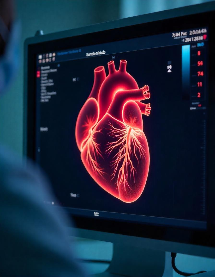
A 2D echocardiogram (2D Echo) is a non-invasive imaging test that uses high-frequency ultrasound waves to create real-time images of the heart. It provides detailed visualization of the heart’s structure, movement, and function, helping in the diagnosis of various cardiac conditions.
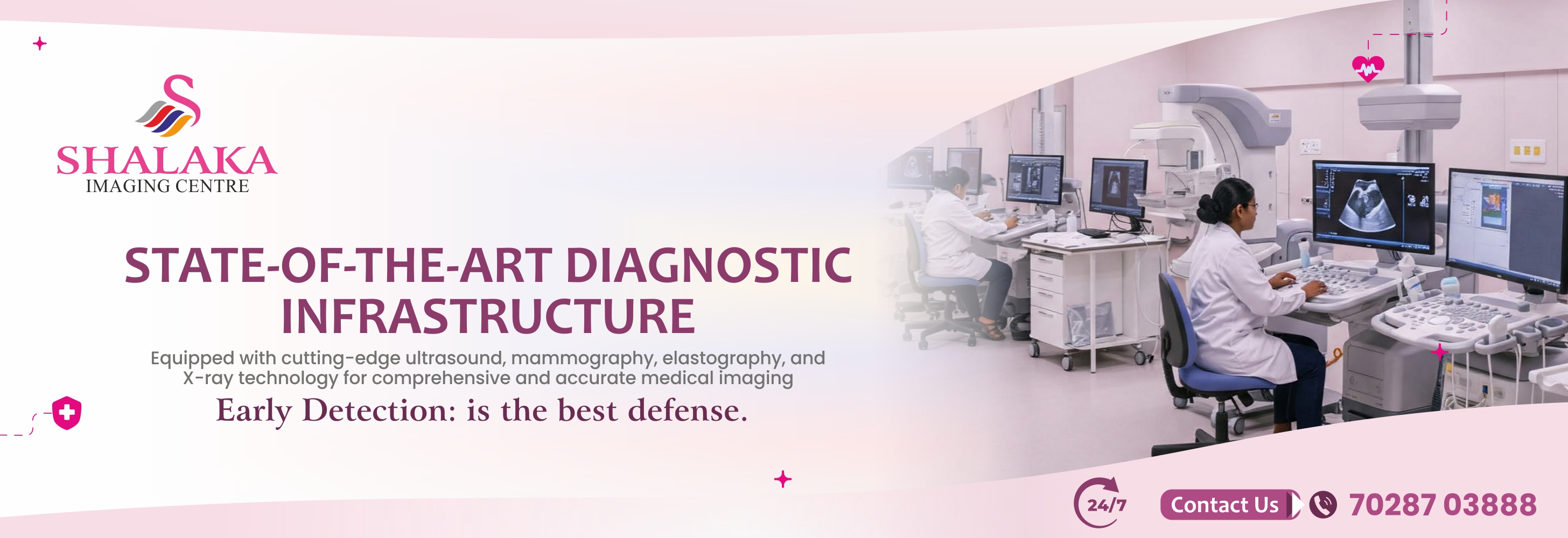
At Shalaka Imaging Center, we offer advanced 2D echocardiography performed by experienced cardiologists and radiologists to assess heart health accurately.
How Does a 2D Echocardiogram Work?
- Ultrasound Transmission – A probe (transducer) placed on the chest emits sound waves that bounce off the heart.
- Image Formation – The returning echoes are processed to create moving images of the heart on a monitor.
- Assessment – The doctor analyzes heart chambers, valves, blood flow, and overall function to detect any abnormalities.
This test is completely safe, painless, and does not involve radiation.
Uses of 2D Echocardiography
A 2D echo test is essential for diagnosing and monitoring various heart conditions, including:
1. Structural and Functional Heart Assessment
- Evaluates heart size and shape, detecting any abnormalities.
- Assesses heart valve function to identify conditions like stenosis or regurgitation.
- Measures heart wall thickness and muscle strength.
2. Diagnosis of Heart Conditions
- Congenital heart defects – Detects structural abnormalities present since birth.
- Cardiomyopathy – Evaluates thickening or weakening of the heart muscle.
- Heart valve diseases – Identifies valve narrowing, leakage, or improper closure.
- Pericardial effusion – Detects fluid buildup around the heart.
- Aneurysms – Identifies bulging or weakened sections of blood vessels.
3. Monitoring Heart Function
- Assesses pumping efficiency in patients with heart failure.
- Helps in pre-surgical evaluation before heart procedures.
- Monitors the effects of medications and treatments on heart performance.
Advantages of 2D Echocardiography
- Non-Invasive & Painless – No injections or surgery required.
- Real-Time Imaging – Provides live visualization of heart movements and function.
- Accurate Diagnosis – Detects heart diseases at an early stage.
- Radiation-Free – Uses ultrasound waves, making it completely safe.
- Quick and Efficient – The procedure takes about 20 to 30 minutes with immediate results.
Why Choose Shalaka Imaging Center for 2D Echocardiography?
- State-of-the-Art Ultrasound Technology – High-resolution imaging for precise diagnosis.
- Expert Cardiologists and Radiologists – Specialists with extensive experience in cardiac imaging.
- Comfortable and Patient-Friendly Care – Ensuring a stress-free and smooth experience.
- Fast and Reliable Reports – Quick test results for timely treatment decisions.